


A honeybee colony’s home is a deliberately stable environment suitable for all sorts of organisms to inhabit, not just bees. The daily stream of foraging bees provides effective transport in and out, and the availability of eggs and larvae, aging adults, stored protein, and carbohydrate is an irresistible attraction for both pathogenic and non-pathogenic opportunists searching for a share.
Have you ever thought about the ecology in a honey bee’s hive? Bees are usually the biggest inhabitants, but they are not the only inhabitants. Occasionally there is the odd reveal when something goes wrong but day-to-day we are blind to most of the space’s tiny occupants. The overwhelming majority of this microcosm does not harm or even interact with the bees, but sometimes some of it does.
A question of scale
If metres and kilogrammes are a reasonable scale to measure human beings with, we need one three orders of magnitude smaller for bees, millimetres and grammes. It’s difficult to imagine life at the scale of a bee. A honey bee say, 15 millimetres long, weighs 160 milligrams. Those ‘big’ (Varroa) mites you see, are about 1.5 to 2 millimetres across. If we measured them using a scale three orders of magnitude smaller still we would use microns (1500-2000µm). Most mites are about half to a quarter of that size, pollen grains range roughly from 5 to 100 microns and trypanosomes (Lotmaria and Crithida) are 5 - 30 microns. AFB bacteria measure 3-5 microns and their spores about a single micron.
Looking at things smaller than a bee that have to be measured in fractions of a millimetre, fractions we call microns, gets tricky. The structures in our eyes we use to detect the light coming from such tiny objects are too big to resolve things smaller than about 100 microns apart, so many of the organisms honey bees live with are too small to see.
Discovering the tools
For a long, long time we have used lenses to magnify small things. The first ‘lenses' were observed in nature, things were seen through a water droplet, or in fish or animal eye lenses. There were natural clear ‘reading stones’, used for magnifying text in the 9th and 10th century (we polished them up a bit first) and as we learnt to work with glass, spectacles and microscopes were developed in the 13th century. Lenses can change the angular incidence of the light from small objects so that they appear larger, but it wasn’t until the 17th century that we started to get both ‘bigger’ and ‘clearer’, the sharpness or ‘resolution’ of the larger image depended mainly on the quality of the lenses improving.
New horizons
Lenses as used in microscopes, good enough to discern something measuring around thousand microns (1mm), were a practical proposition for ‘naturalists’ (people studying Natural History) by the middle of the 17th century. They were a hot topic at the newly created scientific group, the Royal Society in London, and its members were fascinated enough to foster the publication and distribution of Robert Hooke’s Micrographia, which collected the painstakingly detailed, larger-than-life drawings the author had made of everyday subjects, cheese mould, bits of plants, fleas, and so on1. It was the first time images like this had been seen, and they were as impressive and ‘other-worldly’ as images from the James Webb telescope or scanning electron microscopes are today. Even Samuel Pepys, the famous London diarist, had a copy of Micrographia. It was the year 1665 and we could discern the detail in objects we could barely see.
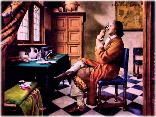
It was a another man, who corresponded with the Royal Society at the time and Hooke’s closest ‘competitor’, that has become known as the ‘father’ of microbiology. Antoni van Leeuwenhoek lived in the Netherlands and was a Delft draper by trade. He was a keen and skilled microscopist who developed his own lens-making techniques and made more than 20 single lens instruments that redefined ‘micro’. He wanted to see thread in fabric apparently.

Ironically Leeuwenhoek never thought of himself as the father-figure of a new scientific discipline, he never published scientific papers or visited the Society, only wrote in Dutch, and refused to share any of his techniques. Some of his specimens collection still remains in the Royal Society archive and can still be examined. A recent investigation (2021) using modern technology2 has shown that at least some of Leeuwenhoek’s best lenes (Hooke never knew this) were made using the method Hooke had developed but never perfected. The lens used was a tiny globule of glass formed by melting and stretching a fine glass rod, and then re-melting the glass strand in a flame.
In 1676 Leeuwenhoek sent the Society the first observations of an organism that had a single cell and was scarcely believed. People had guessed that there were disease transmitting agents that were too small to see a long, long time ago, and had even given them a name - ‘animicules’. Leewenhoek’s meticulous early microscopy had revealed detail at the scale of a single micron, an invisible world discovered 2000 years after it had been imagined.
Revealing the detail
The work to explore and describe these other worlds in the 17th century has interesting parallels with our 21st century images. If you have struggled with your pollen grains even under a modern microscope you’ll remember that the sample preparation matters, lighting and aperture control are often beyond manageable, and focus is finicky. In the 17th century you had to be able to draw (photography is 200 years in the future), and your drawing would be re-constructed by someone else and wood-cut, engraved or etched so that it could be printed. Drawings from Micrographia are composites, made up from many samples and viewing different parts at various angles and focus planes and took days, at best, to create. They were then recreated as prints from etchings in copper plate. Underlying the skill was a desire to reveal the perfection of God’s Creation, Micrographia is both Art and Science3.
In the years that followed we learnt to improve the contrast between one part and another with coloured stains, we got more skilled at managing light using ‘bright field’, ‘dark field’, ‘phase-contrast’ and ‘interference’, even ‘fluorescence’ and ‘confocal’ laser microscopes, but eventually our need to ‘see’ smaller with more detail exceeded the utility of light itself. We have to probe with X-rays, or electrons, and computers compose, ‘false-colour’, and interpret our images; it’s the art of science.
A hive’s microcosm
And what else have we seen living in bee-hives? Depending where you are, in roughly descending size order there are of course beetles (Coleoptera), spiders and pseudoscorpions (Arachnidae), moth larvae (Lepidoptera), earwigs (Dermaptera), silverfish (Thysanura), and ants (Formicidae). Down at the near-impossible-to-see scale, the flies Braula and melaloncha (Diptera) are found. At least 83 mite species (Acari) have been associated with honey bees, both resident and hitchhiking, 13 are fairly common. Even excluding varroa, it wouldn’t be surprising for there to be seven times as many individual mites as bees in a hive, although with near constant doses of mite-lethal ‘varroacides’ who knows. With good light microscopes nematodes (Mermithidae), flagellates (Lotmaria, Crithidia), other protozoa (Gregarines, Amoeba, Variomorpha), fungi (Ascophaera and Bettsia), and algae just come in to view, but for a really detailed picture you need to go beyond light. With electron microscopes, many bacteria, including spiroplasmas, and suite of viruses.
What’s next?
Powered by advancing nanotechnology investment, it’s not a matter of increments of scale anymore. Modern electron microscopes are already examining molecular scales another three orders of magnitude smaller than microns (nanometres, 10-9) researchers can see images of folded proteins less than 10 nanometres across. Scanning Probe Microscopy includes types of microscope that use a fine probe to scan the surface of a specimen with electrons tunnelling between the tip and the sample to create (reconstruct might be a better word) high-resolution, three-dimensional views at the picometre scale (10-12) of atoms. Computers control the microscope and correct, enhance, segregate, and classify multiple images in real time, potentially in living samples. The art in science. We are fast approaching the point where, not only do we see a honey bee virus particle, we can imagine watching the physics of its chemistry.
*This article first appeared in the Apiarist’s Advocate, September issue 50
You can see, and turn the pages on a wonderful copy of ‘Micrographia: or, some Physiological Descriptions of Minute Bodies made by Magnifying Glasses, with Observations and Inquiries thereupon’ digitised by the Royal Society at https://royalsociety.org/blog/2020/07/micrographia-online/
T. Cocquyt, Z. Zhou, J. Plomp, L. van Eijck, Neutron tomography of Van Leeuwenhoek’s microscopes. Sci. Adv. 7, eabf2402 (2021) doi:10.1126/sciadv.abf2402
For a good essay about the truth of images see Patricia Fara’s ‘A microscopic reality tale’, Nature 459, 642–644 (2009). https://doi.org/10.1038/459642a